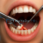
Impacted Wisdom Tooth Extraction
Impacted wisdom tooth extraction is a surgical procedure to remove third molars (wisdom teeth) that have not fully erupted or have emerged in the wrong position. These teeth typically try to erupt between the ages of 17 and 25, but due to limited jaw space, being trapped under the gum, or erupting at an improper angle, they often remain impacted.
What Is an Impacted Wisdom Tooth?
Wisdom teeth are the third molars located at the very back of the upper and lower jaws. When impacted, they can cause the following problems:
-
Jaw pain
-
Gum inflammation
-
Bad breath and altered taste
-
Pressure or damage to adjacent teeth
-
Cyst formation
When Should They Be Removed?
Impacted teeth don’t always show symptoms. However, extraction is recommended in the following cases:
-
Pain or pressure sensation
-
Gum infection
-
Radiographic signs of damage to surrounding tissues
-
Interference with orthodontic treatment
Many recent studies suggest that extraction should be considered as soon as impaction is detected radiographically, even before symptoms appear.
How Is the Extraction Performed?
The extraction is usually carried out under local anesthesia. The surgical steps are as follows:
-
Examination & Imaging: The position of the tooth is evaluated.
-
Anesthesia: The area is numbed for the procedure.
-
Surgical Access: The gum is incised, bone may be removed, and the tooth is extracted in parts if necessary.
-
Suturing & Healing: The area is cleaned and sutured.
Post-Operative Care and Recovery
-
Avoid spitting, rinsing, and using straws during the first 24 hours.
-
Cold compresses may be applied to reduce swelling.
-
Medications prescribed by the dentist should be used regularly.
-
Smoking and alcohol should be avoided for at least 24–48 hours, as they delay healing.
Smoking especially increases the risk of alveolitis (dry socket), a painful inflammation of the bone.
Healing is typically complete within 7 to 10 days.
Impacted Wisdom Teeth: Positions, Nerve Proximity, and Effects on Adjacent Teeth
Common Positions of Impacted Wisdom Teeth
Due to space limitations or deviation in eruption path, impacted wisdom teeth may remain embedded in various angles within the jawbone. The most frequent positions include:
-
Vertical Impaction: The tooth is upright but lacks space to erupt.
-
Mesial (Angled) Impaction: The tooth is angled toward the second molar. This is the most common type.
-
Distal Impaction: The tooth is tilted toward the back of the jaw.
-
Horizontal Impaction: The tooth lies completely sideways and usually requires extraction.
-
Fully Impacted: The tooth is completely covered by gum and bone.
-
Partially Impacted: Part of the tooth has erupted into the mouth, while the rest remains under the gum or bone.
These positions directly affect the surgical approach and complexity of extraction.
Relationship with the Mandibular Nerve and the Need for CBCT
Lower wisdom teeth may be in close proximity to the mandibular nerve, which must be carefully evaluated before extraction. Damage to this nerve can result in:
-
Numbness in the lower lip, chin, or tongue
-
Tingling sensations
-
Long-term or permanent sensory loss
Panoramic X-rays are typically the first step in assessment. However, if nerve involvement is suspected, Cone Beam Computed Tomography (CBCT) should be performed to analyze the three-dimensional relationship between the tooth and the nerve. This allows precise surgical planning and reduces the risk of complications.
Problems Caused to Adjacent Teeth
Impacted wisdom teeth can also negatively affect the adjacent second molars. Especially in mesially impacted teeth:
-
Cavities may form on the distal surface of the second molar. These are often hidden and deep, and may be noticed too late.
-
Periodontal pockets and bone loss may occur.
-
Root resorption of the adjacent tooth can develop.
Such complications may not only threaten the impacted tooth but also lead to the loss of a previously healthy second molar.
Conclusion
Although they may appear harmless, impacted wisdom teeth can cause serious problems involving the jawbone, nerves, and neighboring teeth. Therefore:
-
Their position must be carefully evaluated.
-
If nerve proximity is suspected, CBCT imaging is essential.
-
Risk of cavities in adjacent teeth should be monitored through regular check-ups.


